
Page 34 - Delaware Medical Journal - July/August 2020
P. 34
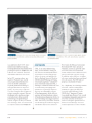
Figure 3-A Admission CT chest, axial image. Note the basilar predominate consolidation, subpleural sparing with ground glass opacities.
Figure 3-B Admission CT chest, coronal image. Again, basilar- predominant consolidation, R>L, with ground glass opacities are demonstrated.
was admitted to the ICU. CT chest demonstrated subpleural sparing, basilar-predominant consolidation with ground glass opacities (Figure 3-A and 3-B), the most commonly described radiographic appearance of EVALI.1
In the ICU, a sputum culture was negative. The patient was started
on Levaquin and azithromycin to
cover for possible community- acquired pneumonia and 60mg IV methylprednisolone for suspected EVALI. Over the course of the next five days, the patient slowly improved and was weaned to room air. She was discharged with a prednisone taper
and instruction to follow up with pulmonology. Her pulmonologist ordered a repeat CT chest, one month after discharge, which was normal with no sequelae demonstrated (Figure 4).
DISCUSSION
EVALI is an acute inhalation lung
������ ���� �� ��������� �������� ��
date. Theories include lipoid-induced and Vitamin E acetate-induced lung injuries. A recent study published in
the New England Journal of Medicine (NEJM) called “Pathology of Vaping- Associated Lung Injury” contained 17 cases of clinically suspected EVALI. Histologic examination showed all cases to include foamy macrophages and pneumocyte vacuolization. However, there was no radiographic evidence
of lipoid pneumonia, which calls into questions utilizing histologic evidence
of lipoidization as diagnostic criteria. As ����� ��� ������� ��� ������� �������� rather than toxicity and caution should be used if utilizing this method of diagnosis until further data has been collected.2
The Centers for Disease Control and Prevention (CDC) have presented Vitamin E acetate as a possible cause of EVALI based on 29 bronchiolar lavage samples from 10 different states that all contained vitamin E acetate.
In addition, other additives, including oils, were analyzed and were not found to be of concern. The CDC did not propose a mechanism of injury.3
So, if lipids may be a histologic marker of EVALI, what are radiographic findings to suggest the diagnosis?
A recently published paper in the NEJM titled “Imaging of Vaping- Associated Lung Disease” lists several overlapping radiographic appearances of EVALI described in literature,
such as eosinophilic pneumonia and diffuse alveolar damage. The most common appearance is that of basilar-
178
Del Med J | July/August 2020 | Vol. 92 | No. 4
