
Page 33 - Delaware Medical Journal - July/August 2020
P. 33
CASE STUDY
INTRODUCTION
Acute inhalation injury is defined as injury to pulmonary epithelium at various levels of the respiratory tract secondary to an inhaled substance. There are many environmental inhaled substances that may cause acute inhalation, which may be passively or actively inhaled. The spectrum of lung injury depends on factors related to the exposure of the inhaled substance, such as the characteristics of the substance (solubility, composition), the amount (dose), and length of exposure (time). Known environmental and workplace exposures such as metals, acids, bases, and solvents are well described in literature with predictable presentations and sequelae.
One new entity, which has caught national headlines recently, is E-cigarette, or vaping, product use- associated lung injury (EVALI).
This disease process is not yet well understood. It has been postulated that accumulation of lipids, such as mineral oils, may be the cause. The theory is based on the fact that vaping cartridges are mixed with a solvent that may be heated up to the point of combustion where there is a phase change to vapor, which may be inhaled. Once inside
the lung alveoli, the contents (nicotine or THC) may be absorbed into the capillaries. But the solvent temperature rapidly declines, changing the phase
of the oil from vapor back to liquid. This liquid oil cannot escape the lung bronchiole tree, and lipid-induced inflammation, lipoid pneumonia, is the result.
CASE PRESENTATION
A 20-year-old female presented
to the hospital with seven days of progressive shortness of breath, fevers
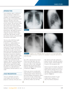
Emergency room radiograph. Note the progression of increased interstitial markings to multifocial airspace opacities. This radiograph is nonspecific and includes a wide differential.
Figure 1
Figure 2
up to 103.6, and post-tussive emesis with no prior lung injury. She was seen in an urgent care facility three days prior, where a chest radiograph demonstrated increased interstitial markings compatible with bronchial inflammation (Figure 1).
Follow-up exam was recommended
with correlation to environmental or inhaled exposure history. Patient was prescribed 40mg oral prednisone, which she took for two days without much improvement, and instructed to follow up with her primary care provider.
Her shortness of breath continued to progress and she eventually developed perioral cyanosis, which prompted her to report to the emergency room.
Upon arrival to the hospital, the patient saturated 86% on room air, low 90s on nasal cannula, and upper 90s on a non- rebreather. A chest radiograph at that time demonstrated multifocal airspace opacities (Figure 2).
Further history elicited 1.5 years of daily, heavy vaping use. CT chest was ordered, and patient
Del Med J | July/August 2020 | Vol. 92 | No. 4
177
