
Page 35 - Delaware Medical Journal - May/June 2019
P. 35
CASE REPORT
Most cases show no evidence of disease recurrence and there have been no reported cases of metastasis.� This report presents
a case of AFST arising in the lower lateral thigh of a young adult male, which was successfully treated with surgical resection, and demonstrated a previously undescribed ������� �������
CASE REPORT
A 20-year-old male was referred to surgical oncology at our institution for evaluation �� � ���� �������� ���� ������� ����
his right lower lateral thigh. The patient initially noticed the mass at least six months prior to presentation, but did not recall any appreciable growth over time. There was no association with leg swelling
or neurologic symptoms. There was no
���������� ����������� �� ���������� ����
medical history.
Magnetic resonance imaging (MRI) revealed an ovoid, well-marginated 5.0 x ��� � ��� �� ���� ������ ��� ������ ������ of the lateral right thigh surrounding
the iliotibial band (Figure 1). The mass demonstrated intermediate T1 signal, which was of slightly greater intensity than muscle, and homogenously hyperintense T2 signal. Post-contrast T1-weighted images revealed homogenous avid enhancement of the mass. Transfascial extension was demonstrated, with portions �� ��� ���� ������� ���� ���������� ��� deep to the iliotibial band. There was
���� �� � ����������� ����� ������� ��� mass and the adjacent vastus intermedius
muscle, with the latter also appearing mildly compressed and displaced.
�������� ���������� ����������� ���
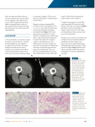
needle aspiration (FNA) and core needle biopsies of the mass were performed (Figure 2). Similar to the preceding MRI, CT images acquired at the time of the biopsy also showed the soft tissue mass to extend across the iliotibial band.
Histology was reviewed at our institution and the Mayo Clinic, and revealed an elaborate proliferation of small, capillary-sized
blood vessels in association with generally bland-appearing stromal cells and a variable amount of collagen (Figure 3). Based on the clinical, radiologic, and histologic features, ��� ��������� �� � ���� ������ ����������� was made.
Figure 2: Axial unenhanced CT images obtained at the time of the biopsy procedure. (a) A soft tissue mass in the lateral thigh has components located both superficial and deep to the iliotibial band. (b) Post-needle placement CT image demonstrates the needle tip to be within the targeted mass.
Figure 3: H&E stains at 40x magnification demonstrate (a) (b) spindle cells without cytologic atypia, set within a myxoid and edematous stroma, with collagen bundles and numerous small, thin- walled, branching blood vessels.
Del Med J
| May/June 2019
| Vol. 91 |
No. 3
131
