
Page 34 - Delaware Medical Journal - May/June 2019
P. 34
Transfascial Soft Tissue Angiofibroma
� C. Eric Gullbrand, DO; Mohammed M. Ali, MD; Eric Montgomery, MD; Gregory Tiesi, MD; Mandip Gakhal, MD
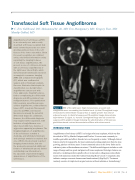
Figure 1: MRI of the right lower thigh demonstrates an ovoid, well- marginated mass surrounding the iliotibial band. (a) Axial T1-weighted image demonstrates intermediate T1 signal that is slightly greater than that of adjacent muscle. (b) Axial fat-suppressed T2-weighted image demonstrates hyperintense T2 signal. (c) Coronal T1-weighted image and (d) coronal fat- suppressed T1-weighted image following the administration of intravenous gadolinium-based contrast demonstrates diffuse avid enhancement.
INTRODUCTION
����������� �� ���� ������� ������ �� � ������ ���� ������ ��������� ����� ��� ����
described in 2012 by Mariño-Enríquez and Fletcher.1 It occurs most commonly in middle-aged adults and affects females twice as frequently as males.1 Although clinical �������� ���� �� �� ����������� ��� ���� ������ ���������� ������� �� ���� �� � ����� growing, painless soft tissue mass.1 It most commonly arises in the lower limbs and in �������� �� ������ �� ������������� �����������1 The differential diagnosis includes a wide range of benign and low-grade malignant soft tissue neoplasms. Histologic features are distinctive, including uniform spindle cells set in myxoid or collagenous extracellular matrix with numerous small, thin-walled, branching blood vessels.1 Cytogenetic data ��������� � ������ ��������� ����������� ������������� ����������������2 Treatment routinely consists of simple local surgical excision without radiation or chemotherapy.�
Angiofibroma of soft tissues (AFST)
is an extremely rare and recently described soft tissue neoplasm that most commonly presents as a slow- growing, painless mass in the soft tissues of the lower extremities. Given its recent recognition as a distinct entity, only limited information exists regarding the imaging features
of soft tissue angiofibromas. We present a case of a 20-year-old male with a soft tissue angiofibroma of
the right lower lateral thigh, which demonstrated transfascial extension on magnetic resonance imaging (MRI) and computed tomography (CT), which was confirmed at
surgery and on histologic analysis. Our case illustrates that despite its classification as a benign tumor, angiofibroma can present with
more aggressive imaging features, further complicating the differential diagnosis and differentiation from malignant tumors. Prior publication lists low-grade myxofibrosarcoma, cellular angiofibroma, solitary fibrous tumor (SFT), low-grade fibromyxoid sarcoma (LGFMS), and myxoid liposarcoma among the potential differential diagnosis candidates. However, the differential diagnosis may require further expansion, with inclusion of angiofibroma even
when faced with more invasive or aggressive-appearing neoplasms. The imaging diagnosis of angiofibroma
of the soft tissues remains a challenge, and ultimately histologic and cytologic analysis is required. Our case affirms the previously reported imaging characteristics, but also
the previously unreported additional feature of transfascial spread, further contributing new information to
the spectrum of potential imaging appearances of angiofibroma of the soft tissues.
130
Del Med J | May/June 2019 | Vol. 91 | No. 3
Abstract
