
Page 33 - Delaware Medical Journal - July/August 2019
P. 33
CASE REPORT
INTRODUCTION
The most widely accepted method of assessing average glycemic control is measurement of hemoglobin A1c (A1c). However, in clinical practice, A1c is really an average and does not represent daily glycemic variability. Patients taking multiple-dose insulin therapy whose A1c falls below 7% tend to have a greater risk of hypoglycemia. However, there are many circumstances under which measured A1c can be falsely low. One of the reasons for variable A1c is red-blood-cell turnover. Anemia or red- cell destruction is known to produce a falsely low A1c.1
Anemia may be found in 40% of patients with colon cancer.2 Type 2 diabetes patients have been shown to have a slight increase in colon cancer risk, especially if they have been obese for many years.3 We report a case of a diabetes patient who had not received routine colon cancer screening and was diagnosed with colon cancer after the discovery of a falsely low A1c.
CASE REPORT
The patient is a 65-year-old male with
history of hypertension, hyperlipidemia, and type 2 diabetes treated with insulin detemir 40 units BID, insulin aspart 20 units TID AC, metformin 1000mg BID, and liraglutide 1.8mg daily, presented with A1c of 5.6% in September 2017. He has had a diagnosis of type 2 diabetes ��� �� ����� ��� ��� ��� ��� ��������� between 7% and 8% for years. In February 2017, patient had A1c of
6.9%, which is close to his usual A1c.
��� �������� �������������� ����������
glucose readings — taken four times daily — correlated with his A1c. There ���� �� ���������� ������� �� ��������� ��� ��� ������� ���������� ������� values from February 2017 to September 2017 (Table 1). Interestingly, patient had not made any dietary changes and there were no changes in his diabetic regimen between February and September.
Thus, the dramatic decrease in A1c was perplexing.
���� ��� ������� ��������� �� ��� �����
visit with A1c of 5.6% in September 2017, he appeared more pale than usual and complete blood count (CBC) and iron panel were ordered to further investigate. A review of his self- monitored blood glucose readings demonstrated no hypoglycemia.
On CBC, patient was found to have microcytic anemia with hemoglobin of
8.3 g/dL (13.3-17.7 g/dL), hematocrit of 31.0% (40-52%), MCV of 66.8 fL (80-100 fL), and MCHC of 26.8% (31.5-36.3%). His iron panel was also ���������� ��� ���� ��������� ������ with Fe of 21 mcg/dL (40-150 mcg/ dL), ferritin of 5 ng/mL (30-400 ng/ mL), transferrin saturation of 4% (15- 55%), and total iron binding capacity (TIBC) of 481 mcg/dL (220-440 mcg/ dL). He did not report any blood in his stools, but given the clinical picture and his age and no prior colonoscopy, he was asked to obtain a colonoscopy. On colonoscopy, he was found to
have mass in the distal transverse
colon, which was found to be a stage 3 adenocarcinoma of colon, and patient received left hemicolectomy, followed by chemotherapy. Patient’s hemoglobin has since then increased to 12.5 g/dL in January 2018. His A1c also increased back to 6.9% and he continues to require insulin for his diabetes (Table 1).
DISCUSSION
In this case, patient was found to have colon cancer after a falsely low A1c was suspected. This case demonstrates the importance of looking at lab work with a certain level of scrutiny and further exploring lab work that does
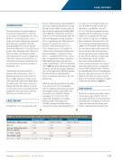
Table 1: Patient Characteristics at Baseline, Presentation, and Treatment of Colon Cancer
February 2017
September 2017
January 2018
Weight (lbs) / BMI (kg/m2)
353.1 / 50.49
354.5 / 50.69
327.2 / 46.78
A1c (%)
6.9
5.6
6.9
Average Fasting Fingerstick Glucose (mg/dL)
144
130
152
Creatinine (mg/dL)
0.80
0.77
0.80
Hemoglobin (g/dL)
-
8.3
12.5
Abbreviations: BMI = body mass index; A1c = hemoglobin A1c
Del Med J | July/August 2019 | Vol. 91 | No. 4
177
