
Page 28 - Delaware Medical Journal - September/October 2020
P. 28
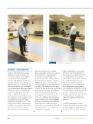
Figure 2 Gait lab test – stance phase.
MATERIALS AND METHODS
A plastic Axis Scientific anatomy-
leg skeleton model (Evanston, IL)
was used to analyze the axial plane measurements and compare the accuracy and reliability among three methods: EOS, CT scan, and gait laboratory analysis. In the beginning, three measurements were performed with each method in the intact original- leg skeleton model to determine the baseline morphology of the model
in the axial plane. Each test was performed at intervals of three weeks. The positioning of the skeleton model was similar to patient positioning during the performance of a CT scan (lying down) and EOS (standing up) test. However, no specific positioning protocol was followed other than
the technicians were instructed to
Figure 3 Gait lab test – swing phase.
position the limb as they would a patient (reproducing the typical patient position). Prior to considering the osteotomy, two parallel pins were placed in each femur and tibia. Axial photographs were made as a reference (Figure 1)� ����� � ��� �����������
was performed in the proximal third of the shaft of the femur and in the distal third of the shaft of the tibia, producing an axial plane deformity of 40 degrees (internal rotation) in the femur and
35 degrees (external rotation) in the tibia. The changes at the osteotomy sites were confirmed with repeat axial images with the pins in place (Figure 1). These osteotomies were fixed with plates and screws and tested again three times for each method, following the same protocol. The results were recorded and compared.
Digital radiographic images using EOS were made from the pelvis to
the foot (just one leg) in both frontal and sagittal planes. The skeleton
bone model was positioned similarly to a weight-bearing position of a patient. One trained technician performed every test and made the three-dimensional reconstruction. Bony landmarks included the femoral head and greater trochanter, posterior contours of the femoral condyles, posterior contour of the proximal tibia, and the malleoli of the ankle joint, which were identified in the usual manner.1,6
Computed tomography (Siemens Somaton Definition Flash Dual Energy, Siemens Healthcare Diagnostics, Inc., ���������� ��� ��� ���� ���������
220
Del Med J | September/October 2020 | Vol. 92 | No. 5
