
Page 29 - Delaware Medical Journal - July/August 2019
P. 29
CASE REPORT
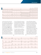
ventricular tachycardia less likely. Her other vital signs were stable, and labs were unremarkable. Initial treatment with vagal maneuvers, including REVERT, and adenosine were both successful in temporarily converting her out of the SVT, but only for a few minutes. Her post-conversion EKG (EKG 2) showed
a sinus dysrhythmia with intermittent junctional beats. Subsequent attempts to control the supraventricular tachycardia (SVT) with longer-acting nodal blockers were ultimately unsuccessful. The patient’s blood pressure did not tolerate intravenous metoprolol, and a diltiazem drip was only able to slow the rate of the
SVT to 140-150 bpm before the patient would become hypotensive. Throughout these interventions, the patient would spontaneously convert in and out of SVT, and with the exception of feeling the palpitations, she remained asymptomatic. The patient was admitted to the cardiology service for refractory SVT.
Over the next three days, cardiology was unable to suppress the SVT despite multiple antidysrhythmic medications. The patient’s blood pressure would
not tolerate the doses of beta-blockers or calcium channel blockers needed
to maintain sinus rhythm, and rhythm
������� ���� ��������� ��� �������
was only transient. On day three of her admission, an echocardiogram showed a left ventricular ejection fraction (LVEF) of 20-25%. This was attributed to a tachycardia-induced cardiomyopathy and prompted the decision to take her
to the electrophysiology (EP) lab for mapping and ablation. EP studies showed a retrograde-only accessory pathway with no observed spontaneous or inducible anterograde conduction. This is formally termed a concealed accessory pathway, ����� �� ������ �� � ������� ���� ���� conducts retrogradely and does not affect the EKG pattern during sinus rhythm.
Del Med J | July/August 2019 | Vol. 91 | No. 4
173
EKG 2 EKG 1
