
Page 36 - Delaware Medical Journal - July-August 2018
P. 36
will demonstrate a rare etiology of ODS in a normonatremic, but hyperglycemic patient.
Case Presentation
A 66-year old male with a past medical history of insulin- dependent diabetes mellitus and osteomyelitis requiring above- the-knee amputation presented to the Wilmington ED after a suspected seizure at the patient’s homeless shelter. After transfer to Christiana Care for admission, the patient was nonverbal and could only follow commands intermittently. Neurological exam ������ �� ����� ������� �� ������� ������� ��������� �� ���������� The patient had a history of recurrent ED visits for uncontrolled hyperglycemia and an HbA1C of 12.9.
Initial labs showed glucose of 597 and sodium of 128. Alcohol level and urine drug screen were negative. Urinalysis and chest x-ray were unremarkable. The patient was initially treated with insulin, �� ������ ��������� ������� �������������� ��� ������������
Throughout the patient’s stay in the hospital, he remained largely non-verbal, responding inappropriately and with one-word answers. The encephalopathy work-up revealed a normal TSH, ammonia, folate, B12, ANA, and ANCAs. EEG during a suspected episode
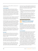
of shaking showed no seizure activity. An LP and TTE showed no signs of infection. Finally, head MRI showed restricted diffusion noted throughout a large portion of the pons in a symmetric distribution bilaterally, concerning for osmotic demyelination.
Blood cultures eventually grew Proteus Mirabilis and a repeat urinalysis was positive for leukocyte esterase and nitrite. A course of antibiotics was completed and there was some improvement in the patient’s mental status and ability to communicate over the following week.
Discussion
This case demonstrates that ODS may be present in a patient with no alteration in their serum sodium, but with hyperosmolar hyperglycemia instead. This patient’s sodium, corrected for �������� ��� ��� ��� ���� ��� �� ���� ��� ������� ������ disturbances of ODS.
Given this patient’s extensive history of non-compliance with his diabetes medications and recurrent hospital visits, it is likely that his ������� ������ ���� ����� �� ���� ����� ���� ���� �������� ����� where ODS was also caused by hyperglycemia, with glucose levels at 4923, 5241, and 6004, in the setting of normonatremia.
�������������� �� ��� ����� ����� �� ������������ ��� ������
by hyperglycemia, all patients achieved at least partial resolution of their symptoms within one month and hopefully the small improvement noted in this patient will follow a similar trend.
Given the increase in our diabetic population throughout the United States,2 this is a condition that will likely increase in incidence; it is important to have a high index of suspicion for ODS when a diabetic patient presents with atypical neurologic symptoms.
REFERENCES
1. Burns JD, Kosa SC, Wijdicks EF. 2009. Central Pontine Myelinolysis in a Patient with Hyperosmolar Hyperglycemia and Consistently Normal Serum Sodium. Neurocritical Care 11 251–254. ()10.1007/s12028- 009-9241-9
2. Rowley WR, Bezold C, Arikan Y, Byrne E, Krohe S. Diabetes 2030: Insights from Yesterday, Today, and Future Trends. Population Health Management. 2017;20(1):6-12. doi:10.1089/pop.2015.0181.
3. Saini M, Mamauag MJ, Singh R. 2015. Central Pontine Myelinolysis: A Rare Presentation Secondary to hyperglycemia. Singapore Medical Journal 56 e71–e73. ()10.11622/smedj.2015065
4. Talluri S, Charumathi R, Khan M, Kissell K. Atypical Presentation of Central Pontine Myelinolysis in Hyperglycemia. Endocrinology, Diabetes & Metabolism Case Reports. 2017:17-0064. doi:10.1530/EDM-17-0064.
STUDENT POSTER SECOND PRIZE
Euglycemic DKA in a Type 2 Diabetic Using Sodium-Glucose Co-Transporter-2 Inhibitor By Ruchika Bhargav, MS4 and Amy Wachter MD
Introduction
Sodium-glucose co-transporter-2 (SGLT-2) inhibitors comprise a new class of oral medications for the treatment of type 2 diabetes mellitus. They effectively lower blood glucose levels through increased urinary excretion of glucose. However, there have been an increasing number of cases recently of euglycemic diabetic ketoacidosis (DKA) in type 2 diabetic patients treated with SGLT- 2 inhibitors.
Case Presentation
A 64-year-old Caucasian female with a history of type 2 diabetes mellitus, Charcot arthropathy of right foot, and tobacco abuse presented to Christiana Hospital with a two-day history of worsening swelling and redness of her right foot along with complaints of nausea and several episodes of vomiting. Her vital signs upon admission included the following: Temperature – 38.2°C, Heart Rate – 120, Respiratory Rate – 18, Blood Pressure – 117/54, and SpO2 – 97%. Physical examination was notable
for an area of erythema on the medial aspect of the right foot. Laboratory studies revealed a mild leukocytosis of 12.4 K/uL with a 6% bandemia, an anion gap of 23, venous pH of 7.15, bicarbonate level of 9.7, a lactic acid level of 1.5, a serum beta-hydroxybutyrate level of 6.7, and a blood glucose level of 174. A diagnosis of euglycemic diabetic ketoacidosis was made. The patient was given 1 liter of normal saline and transferred to Wilmington Hospital ��� ��� ������� ���� ��� �������� �� ����� ��� ������������
was discontinued. The patient’s symptoms and metabolic acidosis ������������ �������� ���� ��� ���� ������� �����
208
Del Med J | July/August 2018 | Vol. 90 | No. 6
