
Page 22 - Delaware Medical Journal - January 2018
P. 22
�������������������������������������������������������������
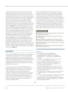
her better hearing ear. In 2016, the patient’s left ear was then implanted with complete electrode insertion. She experienced a hearing improvement on the left as well, and currently possesses functional hearing on both sides with 92 percent word recognition in sentences in quiet. Prior to the 2015 cochlear implantation
in the right ear, the patient underwent a spiral-CT temporal
bone study utilizing 0.625 mm slice thickness. These images demonstrated extensive lucency in the immediate vicinity of both the cochlea and vestibule, consistent with the patient’s history of otosclerosis (Figure 1). Two months following the CT scan, the patient underwent an MRI study of the temporal bones. These images were acquired utilizing a 1.5T MR scanner, and included thin section images through the temporal bones. A thin-section 3D, heavily T2 weighted GRE Constructive Interference in
the Steady State sequence (CISS), demonstrated marked signal abnormality surrounding the cochlea. This area of abnormal signal corresponded to the area of abnormal perichoclear
lucency demonstrated on the CT scan. Remarkably intense �������������������������������������������������������������� and perilabyrinthine distributions, corresponding to the signal abnormality on MRI and abnormal lucency on the CT scan. Pathologic enhancement is very seldom observed on MR imaging of patients with this disorder and, when present, it might suggest an active or aggressive phase of the pathologic processes leading to otosclerosis (Figure 2).
DISCUSSION
Cross-sectional imaging plays an important role in the diagnosis and management of otosclerosis. Due to the typical histologic changes of the middle and inner ear structures during disease ���������������������������������������������������������������� in patients with documented hearing loss. It additionally may aid in diagnosis by eliminating other potential causes for hearing loss including acoustic neuromas, cholesteatomas, a congenital malformations, or infection.
This case illustrates the critical role that imaging plays in
��������������������������������������������������������������
being evaluated for sensorineural hearing loss. Although conductive hearing loss occurs primarily in fenestral otosclerosis, the patient may also present with sensorineural or a mixed type hearing loss if the osseous abnormality extends to the cochlea. The patient presented herein experienced sensorineural hearing loss secondary to cochlear otosclerosis, with almost complete destruction of both otic capsules, as shown by her imaging studies.
The preferred imaging protocol for evaluating otosclerosis is
a non-enhanced spiral CT of the temporal bone, secondary to
its ability to detect small abnormalities of the bony labyrinth ��������������������������������������������������������������� preoperative surgical planning, as well as aid in the prediction of successful surgical outcomes. It is necessary for the CT protocol to be obtained with a slice thickness of 0.5-0.625 mm with an increment of 0.3 mm to allow for the ability to obtain high quality multiplanar reformatted images.7 If MR imaging is requested,
a T1 contrast enhanced sequence should be obtained in order to identify enhancing foci from the acute phases of otosclerosis. A high resolution T2 MR imaging alone may miss otosclerosis.5
CONTRIBUTING AUTHORS
■ KATIE TAYLOR, DO is a fourth year diagnostic radiology resident at Christiana Care Health System in Newark, Del.
■ ALBERTOIAIA,MDisChiefofNeuroradiologyatChristianaCareHealth System in Newark, Del.
■ MICHAELTEIXIDO,MDisanOtolaryngologistatChristianaCareHealth System in Newark, Del. and Thomas Jefferson University in Philadelphia, Penn.
■ FRANCESCOAGNELLO,MDisadiagnosticradiologyresidentattheUniversity of Palermo in Palermo, Italy.
■ GIANVINCENZOSPARACIA,MDisonthediagnosticradiologyfacultyatthe University of Palermo in Palermo, Italy.
REFERENCES
1. Juliano AF, Ginat DT, Moonis G. Imaging Review of
the Temporal Bone: Part II. Traumatic, Postoperative,
and Noninflammatory Nonneoplastic Conditions. Radiology. 2015;276(3):655-72. doi:10.1148/radiol.2015140800. PMID: 26302389
2. Moumoulidis I, Axon P, Baguley D, et al. A review on the genetics of otosclerosis. ClinOtolaryngol 2007;32:239–47
3. Whetstone J, Nguyen A, Nguyen-Huynh A, Hamilton BE. Surgical and clinical confirmation of temporal bone CT findings in patients with otosclerosis with failed stapes surgery. AJNR Am J Neuroradiol 2014;35(6):1195–1201
4. Declau F, van Spaendonck M, Timmermans JP, Michaels L, Liang
J, Qiu JP, van de Heyning P. Prevalence of histologic otosclerosis: an unbiased temporal bone study in Caucasians. Adv Otorhinolaryngol. 2007;65:6-16. PMID: 17245017
5. Swartz NG, Berger AS. Fenestral and cochlear otosclerosis: computed tomographicevaluation. Am J Otol. 1985 Nov;6(6):476-81. PMID: 4073255.
6. Lee TC, Aviv RI, Chen JM, Nedzelski JM, Fox AJ, Symons SP. CT Grading of otosclerosis. AJNR Am J Neuroradiol. 2009; 30:1435–1439
7. Curtin HD. Imaging of Conductive Hearing Loss With a Normal Tympanic Membrane. AJR Am J Roentgenol. 2016;206(1):49-56. doi: 10.2214/ AJR.15.15060. PMID: 26491893.
22
Del Med J | January 2018 | Vol. 90 | No. 1
