
Page 24 - Delaware Medical Journal - February 2017
P. 24
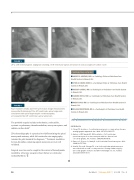
ABC
FIGURE 5
(A-C) Left ventriculogram angiogram showing a left ventricular apical aneurysm in various stages of cardiac cycle.
FIGURE 6
Four chamber steady state free precession image demonstrates myocardial thickening of the left ventricular apical segments, consistent with apical hypertrophic cardiomyopathy, accompanied by left ventricular apical aneurysm.
The probable sequela includes arrhythmias, endocarditis, systemic or pulmonary thromboembolism, aneurysm rupture, and sudden cardiac death.4
2D echocardiography is a practical tool differentiating the apical aneurysmal anatomy, while left ventricular cine angiography remains the gold standard for diagnosis.1-3 Treatment modalities for non-thrombus containing apical aneurysms are not well validated.
Surgical resection can be sought in the context of hemodynamic instability, refractory congestive heart failure or ventricular tachyarrhythmia.2
CONTRIBUTING AUTHORS
■ AHMED S. ABUZAID, MD is a Cardiology Fellow at Christiana Care Health System in Newark, Del.
■ SYED ALI HAMID, MBBS is a Cardiology Fellow at Christiana Care Health System in Newark, Del.
■ MANDIP GAKHAL, MD is a Radiologist at Christiana Care Health System in Newark, Del.
■ SANDRA WEISS, MD is a Cardiologist at Christiana Care Health System in Newark, Del.
■ ROBIN HORN, MD is a Cardiologist at Christiana Care Health System in Newark, Del.
■ WILLIAM WEINTRAUB, MD is a Cardiologist at Christiana Care Health System in Newark, Del.
REFERENCES
1. 2.
3. 4.
Cheng TO. Incidence of ventricular aneurysm in coronary artery disease, an angiographic appraisal. Am J Med. 1971;50:340-355.
Gatewood RP, Nanda NC. Differentiation of left ventricular pseudoaneurysm from true aneurysm with two dimensional echocardiography. Am J Cardiol. 1980;46:869-878.
Frances C, Romero A, Grady D. Left Ventricular Pseudoaneurysm. JACC. 1998;32:557-561.
Xiea M, Zhoua H, Chenga TO, et al. Left ventricular apical aneurysm associated with normal coronary arteries following cardiac surgery: Echocardiographic features and differential diagnosis. Int J Cardiol. 2013;168:3665-3670.
56
Del Med J | February 2017 | Vol. 89 | No. 2
